Saturday, May 23, 2009

Jam Kerja Organ Tubuh

Hours Body Organ

Below is a glimpse of working hours, our body's organs and there is little description about what we should do in the hours to maintain our health. Hopefully you can help.

Lambung
Hours 07.00 - 09.00
Hour picket flank organs are strong, it's breakfast for the establishment of the energy body throughout the day. Drinking juice or herb should before breakfast, so that the stomach is empty of useful substances soon be merged body.

Spleen
Hours 09.00 - 11.00
Hours picket strong organ spleen, in mentransportasi liquid nutrition for energy growth. When the hours this sleepy, it means the spleen is weak. Reduce consumption of sugar, fat, oil and animal protein.

Heart
Hours 11:00 - 13:00
Hours picket strong heart organ, must rest, avoid heat and physical manner, ambition and emotional disturbances, especially in people with blood vessel.

Heart
Hours 13:00 - 15:00
Hour picket organ weak heart, when people sleep, gather in the blood red heart and organ regeneration process occurs cells hearts. Strong function of the heart when the body strong to prevent any disease.

Lungs
Hours 15:00 - 17:00
Hour picket organ lungs weak, needed rest, sleep for the discharge of toxic and the process of the formation of energy consumption

Kidney
Hours 17:00 - 19:00
Hours picket strong organ kidney, should be used to study the process due to the formation of bone marrow and the brain and intellect.

Lambung
19.00 - 21.00
Hour picket organ weak stomach should not consume food which is difficult or long digested digested or better is to stop eating

Spleen
Hours 21:00 - 23:00
Hour picket organ spleen is weak, the process occurs and the discharge of toxic spleen cell regeneration process. Should rest while listening to music that calms the soul, to enhance immunity.

Heart
Hours 23:00 to 01:00
Hour picket organ weak heart. Bed rest should have been, if still working or begadang can weaken the heart function.

Heart
Hours 01.00 - 03.00
Hour picket organ heart strong. There was a process of toxic disposal / waste of body metabolism. If there is any interference in the function of heart reflected the dirt and disruption. If there is any injury will be felt in pain.

Lungs
Hours 03.00 - 05.00
Hour picket organ lungs strong, the process of waste disposal occurred / organ toxicity in the lungs, occurs when coughing, sneezing, sneezing and sweating indicates the existence of interference lung function. We recommend to use it to get a breath of energy tuberculosis healthy and strong.

Colon
Hours 05.00 - 07.00
Hours picket colon powerful organ, it is regularly biasakan CHAPTER

====================================================================

Versi Indonesia


Jam Kerja Organ Tubuh

Di bawah ini adalah sekilas tentang jam kerja organ tubuh kita dan ada sedikit deskripsi tentang apa yang sebaiknya kita lakukan pada jam-jam tersebut untuk menjaga kesehatan kita. Semoga dapat membantu.

LAMBUNG
Jam 07.00 - 09.00
Jam piket organ lambung sedang kuat, sebaiknya makan pagi untuk proses pembentukan energi tubuh sepanjang hari. Minum jus atau ramuan sebaiknya sebelum sarapan pagi, perut masih kosong sehingga zat yang berguna segera terserap tubuh.

LIMPA
Jam 09.00 - 11.00
Jam piket organ limpa kuat, dalam mentransportasi cairan nutrisi untuk energi pertumbuhan. Bila pada jam-jam ini mengantuk, berarti fungsi limpa lemah. Kurangi konsumsi gula, lemak, minyak dan protein hewani.

JANTUNG
Jam 11.00 - 13.00
Jam piket organ jantung kuat, harus istirahat, hindari panas dan olah fisik, ambisi dan emosi terutama pada penderita gangguan pembuluh darah.

HATI
Jam 13.00 - 15.00
Jam piket organ hati lemah, bila orang tidur, darah merah berkumpul dalam organ hati dan terjadi proses regenerasi sel-sel hati. Apabila fungsi hati kuat maka tubuh kuat untuk menangkal semua penyakit.

PARU-PARU
Jam 15.00 - 17.00
Jam piket organ paru-paru lemah, diperlukan istirahat, tidur untuk proses pembuangan racun dan proses pembentukan energi paru-paru

GINJAL
Jam 17.00 - 19.00
Jam piket organ ginjal kuat, sebaiknya digunakan untuk belajar karena terjadi proses pembentukan sumsum tulang dan otak serta kecerdasan.

LAMBUNG
Jam 19.00 - 21.00
Jam piket organ lambung lemah sebaiknya tidak mengkonsumsi makan yang sulit dicerna atau lama dicerna atau lebih baik sudah berhenti makan

LIMPA
Jam 21.00 - 23.00
Jam piket organ limpa lemah, terjadi proses pembuangan racun dan proses regenerasi sel limpa. Sebaiknya istirahat sambil mendengarkan musik yang menenangkan jiwa, untuk meningkatkan imunitas.

JANTUNG
Jam 23.00 - 01.00
Jam piket organ jantung lemah. Sebaiknya sudah beristirahat tidur, apabila masih terus bekerja atau begadang dapat melemahkan fungsi jantung.

HATI
Jam 01.00 - 03.00
Jam piket organ hati kuat. Terjadi proses pembuangan racun/limbah hasil metabolisme tubuh. Apabila ada gangguan fungsi hati tercermin pada kotoran dan gangguan mata. Apabila ada luka dalam akan terasa nyeri.

PARU-PARU
Jam 03.00 - 05.00
Jam piket organ paru-paru kuat, terjadi proses pembuangan limbah/racun pada organ paru-paru, apabila terjadi batuk, bersin-bersin dan berkeringat menandakan adanya gangguan fungsi paru-paru. Sebaiknya digunakan untuk olah nafas untuk mendapatkan energi paru yang sehat dan kuat.

USUS BESAR
Jam 05.00 - 07.00
Jam piket organ usus besar kuat, sebaiknya biasakan BAB secara teratur
Fungsi Vital Organ-Organ Tubuh Saat Hamil

Vital Organ Function-When Pregnant Body Organs


Blood
Towards the end of pregnancy, blood volume increased gradually until reaching 45%. Uterus and other organs in the body need more blood. Breast gum and also receive additional blood. Bone marrow becomes more active in producing blood cells red. After the baby is born, your body must produce more blood to replace blood lost during the process of childbirth.

Heart
Your heart must work harder to pump more blood throughout the body. To do this extra task, the heart grow big and detaknya increase between 10 to 15 times per minute.

Lung-Lung
Lungs must work harder in order to get enough blood oxygen. Hiruplah fresh air as much as possible and exercise so that rajinlah lungs to function properly.

Kidney
Second function of your kidneys filter and clean the blood. During pregnancy, the kidneys are working harder and better to clean up more blood in your body.

Bladder
Work your bladder akan disturbed by contraction in the uterus next to it. Frequency of waste water will be small. This is perhaps disrupt, but not to reduce the drink because you need lots of liquids.

Joint
Bone-associated bone joint, ditopang, and strengthened by a network of tough muscle called ligamen. Hormones of pregnancy soften ligamen especially around the pelvis to become more flexible so. Therefore, the joint-pelvic joints to be flexible during the process of childbirth, so the baby can be born more easily.
Sore back towards the end of pregnancy may be caused because of the increased kelenturan and pelunakan ligamen around the bone pinggul. Ask the doctor about your bone pinggul exercises that can help avoid back pain.
Kelenturan ligamen-ligamen on the pregnancy can cause pain down the back, foot, and toe. Gymnastics, the right shoes, and massage can help them. Body posture is also very much a role in reducing pain.

========================================

Versi Indonesia

Fungsi Vital Organ-organ Tubuh Saat Hamil


Darah

Menjelang akhir kehamilan, volume darah meningkat secara bertahap sampai mencapai 45%. Uterus dan organ-organ lain dalam tubuh membutuhkan lebih banyak darah. Payudara dan gusi juga menerima tambahan darah. Sumsum tulang menjadi lebih aktif memproduksi sel-sel darah merah. Setelah bayi lahir, tubuh anda harus memproduksi darah lebih banyak untuk mengganti darah yang hilang pada saat proses persalinan.

Jantung

Jantung anda harus bekerja lebih keras untuk memompa darah lebih banyak ke seluruh tubuh. Untuk melakukan tugas ekstra ini, jantung bertambah besar dan detaknya meningkat antara 10 sampai 15 kali per menit.

Paru-Paru

Paru-paru harus bekerja lebih keras agar darah mendapatkan cukup oksigen. Hiruplah udara segar sebanyak mungkin dan rajinlah berolahraga agar paru-paru dapat berfungsi dengan baik.

Ginjal

Kedua ginjal anda berfungsi menyaring dan membersihkan darah. Pada masa kehamilan, kedua ginjal tersebut bekerja lebih keras dan lebih baik untuk membersihkan darah lebih banyak dalam tubuh anda.

Kandung Kemih

Kerja kandung kemih anda akan terganggu oleh kontraksi uterus di sebelahnya. Frekuensi buang air kecil anda akan bertambah. Hal ini barangkali mengganggu, tetapi jangan sampai mengurangi minum karena anda memerlukan banyak cairan.

Persendian

Tulang-tulang persendian dihubungkan, ditopang, dan diperkuat oleh jaringan otot liat yang dinamakan ligamen. Hormon-hormon kehamilan memperlunak ligamen terutama yang disekitar panggul sehingga menjadi lebih lentur. Oleh karena itu, sendi-sendi panggul menjadi fleksibel pada saat proses persalinan sehingga bayi dapat lahir dengan lebih mudah.

Nyeri punggung menjelang akhir masa hamil dapat disebabkan karena peningkatan kelenturan dan pelunakan ligamen di sekitar tulang pinggul. Tanyalah kepada dokter pribadi anda tentang senam tulang pinggul yang dapat membantu menghindarkan nyeri punggung.

Kelenturan ligamen-ligamen lain pada masa kehamilan dapat menimbulkan nyeri dibagian punggung bawah, kaki, dan ujung kaki. Senam, sepatu yang tepat, dan pijatan dapat membantu mengatasinya. Sikap tubuh juga sangat berperan dalam mengurangi nyeri.

Sistem Pernapasan Pada Manusia

The Human Respiratory System


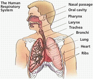
Tools respiratory function include air containing oxygen and removing the air containing carbon dioxide and water vapor.
Respiratory process that is the goal to obtain energy. Breathing events occur in the energy release
Man on the respiratory system consists of
o nose
o faring
o trakea
o bronkus
o bronkiouls
o lungs


Cavity Nose
Cavity on the surface of the nose, there are hair-fine hair and mucous that act to filter the air into the nose cavity.


Base of the throat
Base of the throat organized by some form of cartilage jakun. Base of the throat can be closed by valve base of the throat (epiglotis). At the time of ingest food, close the valve is at the base of the throat and breathing time to open the card. At the base of the throat there is a membrane that will vibrate the sound when there is air from the lungs, for example, at the time we talk.


Trachea
Trachea (trakea) located in the front of the esophagus. In the thoracic cavity, trachea fork bronchus (bronkus). In the lung, bronchus ramify into a very small channel called bronkiolus. Edge bronkiolus form small bubbles, called bubble lungs (alveolus).


Lungs
Lungs are located in the thoracic cavity. Thoracic cavity and the stomach is restricted by siuatu divider called diaphragm. Lungs are the two lungs and the right of the left lung. Right lung consists of three gelambir (lobus) gelambir namely top, middle and gelambir down gelambir. While the left lung consists of two gelambir the gelambir up and down gelambir. Lungs covered by a membrane lung (pleura).
Alveolus in the lungs is very much in number, approximately 300 million alveolus. The entire surface of the alveolus width estimated 100 times greater than the surface of the body. Alveolus dikekelingi-tube capillary blood vessel.


Gas exchange in Alveolus
Oxygen required for oxidation is taken from the air that we hirup at the time we breathe. At the time breathing the air entering through the channel and respiratory akhirnyan into the alveolus. That there is oxygen in the alveolus berdifusi penetrate the cell wall of alveolus. Eventually into the blood vessel, and bound by the hemoglobin in the blood become oksihemoglobin. Further distributed by the blood throughout the body.
Oksigennya released into the body cells back into so oksihemoglobin hemoglobin. Carbon dioxide produced from the respiratory transported by blood through the vein until the end of the alveolus from alveolus of carbon dioxide released through the respiratory channel in the time we issue a breath.
Thus, the exchange occurs in the alveolus oxygen gas that is incoming and outgoing karnbondioksida.


The process of respiratory
Breathing process that includes two interesting breath or respiratory air to enter and remove the air, the breath or respiratory issue. Interesting breath is called inspiration and the breath is called ekspirasi issue.
Interesting at the time of breath, muscle berkontraksi diaphragm. The diaphragm position over the curve is now so straight into the chest cavity. This is called respiratory stomach. Along with diaphragm muscle contraction, muscle-rib muscles also berkontraksi so that the chest cavity. This is called respiratory chest.
Due to the thoracic cavity, the pressure in the thoracic cavity to be reduced, so that the incoming air from outside through the nose, next through the respiratory channel air into the lungs, so the lungs inflate.
Expenditure caused by the breath melemasnya muscles and diaphragm musculature and rib berkontraksinya also assisted with the muscle belly. Diaphragm to be curved up, rib-bone down to the bottom and to move in the direction, consequently chest cavity so that the pressure decline in the thoracic cavity increases. With the increase of pressure in the thoracic cavity, the air inside the lungs out through respiratory channels.


Capacity Lung tuberculosis
The incoming air out the lungs at the time doing normal respiratory air is called respiratory (tidal air). Air volume in the adult respiratory approximately 500 nl. After we do a normal inspiration, we can still attract deep breath-in. Air can enter after a normal inspiration called complementary air, more or less volume 1500 ml. Once we do ekspirasi usual, we can still blow away the breath with a vengeance. Air that can be issued after the regular ekspirasi called air suplementer, volume approximately 1500 ml.
Although we spent the breath from the lungs with a flat-out turns in the lungs are still the air is called air residue. Volume of air residues more or less in 1500 ml. The number of respiratory air volume, air, complementary, and the air suplementer called the vital capacity of lungs.


====================================================================

Versi Indonesia

Sistem Pernafasan Pada Manusia


Alat-alat pernapasan berfungsi memasukkan udara yang mengandung oksigen dan mengeluarkan udara yang mengandung karbon dioksida dan uap air.
Tujuan proses pernapasan yaitu untuk memperoleh energi. Pada peristiwa bernapas terjadi pelepasan energi

Sistem Pernapasan pada Manusia terdiri atas

o hidung

o faring

o trakea

o bronkus

o bronkiouls

o paru-paru


Pernafasan-manusia


Rongga Hidung

Pada permukaan rongga hidung terdapat rambut-rambut halus dan selaput lendir yang berfungsi untuk menyaring udara yang masuk ke dalam rongga hidung.

Pangkal Tenggorok

Pangkal tenggorok disusun oleh beberapa tulang rawan yang membentuk jakun. Pangkal tenggorok dapat ditutup oleh katup pangkal tenggorok (epiglotis). Pada waktu menelan makanan, katup tersebut menutup pangkal tenggorok dan pada waktu bernapas katu membuka. Pada pangkal tenggorok terdapat selaput suara yang akan bergetar bila ada udara dari paru-paru, misalnya pada waktu kita bicara.

Batang tenggorok

Batang tenggorok (trakea) terletak di sebelah depan kerongkongan. Di dalam rongga dada, batang tenggorok bercabang menjadi dua cabang tenggorok (bronkus). Di dalam paru-paru, cabang tenggorok bercabang-cabang lagi menjadi saluran yang sangat kecil disebut bronkiolus. Ujung bronkiolus berupa gelembung kecil yang disebut gelembung paru-paru (alveolus).

Paru-paru
Paru-paru terletak di dalam rongga dada. Rongga dada dan perut dibatasi oleh siuatu sekat disebut diafragma. Paru-paru ada dua buah yaitu paru-paru kanan dan paru-paru kiri. Paru-paru kanan terdiri atas tiga gelambir (lobus) yaitu gelambir atas, gelambir tengah dan gelambir bawah. Sedangkan paru-paru kiri terdiri atas dua gelambir yaitu gelambir atas dan gelambir bawah. Paru-paru diselimuti oleh suatu selaput paru-paru (pleura).
Alveolus dalam paru-paru jumlahnya sangat banyak, lebih kurang 300 juta alveolus. Luas permukaan seluruh alveolus diperkirakan 100 kali lebih besar daripada permukaan tubuh. Alveolus dikekelingi pembuluh-pembuluh kapiler darah.

Pertukaran Gas dalam Alveolus

Oksigen yang diperlukan untuk oksidasi diambil dari udara yang kita hirup pada waktu kita bernapas. Pada waktu bernapas udara masuk melalu saluran pernapasan dan akhirnyan masuk ke dalam alveolus. Oksigen yang terdapat dalam alveolus berdifusi menembus dinding sel alveolus. Akhirnya masuk ke dalam pembuluh darah dan diikat oleh hemoglobin yang terdapat dalam darah menjadi oksihemoglobin. Selanjutnya diedarkan oleh darah ke seluruh tubuh.

Oksigennya dilepaskan ke dalam sel-sel tubuh sehingga oksihemoglobin kembali menjadi hemoglobin. Karbondioksida yang dihasilkan dari pernapasan diangkut oleh darah melalui pembuluh darah yang akhirnya sampai pada alveolus Dari alveolus karbon dioksida dikeluarkan melalui saluran pernapasan pada waktu kita mengeluarkan napas.

Dengan demikian dalam alveolus terjadi pertukaran gas yaitu oksigen masuk dan karnbondioksida keluar.

Proses Pernapasan

Bernapas meliputi dua proses yaitu menarik napas atau memasukkan udara pernapasan dan mengeluarkan napas atau mengeluarkan udara pernapasan. Menarik napas disebut inspirasi dan mengeluarkan napas disebut ekspirasi.
Pada waktu menarik napas, otot diafragma berkontraksi. Semula kedudukan diafragma melengkung keatas sekarang menjadi lurus sehingga rongga dada menjadi mengembang. Hal ini disebut pernapasan perut. Bersamaan dengan kontraksi otot diafragma, otot-otot tulang rusuk juga berkontraksi sehingga rongga dada mengembang. Hal ini disebut pernapasan dada.

Akibat mengembangnya rongga dada, maka tekanan dalam rongga dada menjadi berkurang, sehingga udara dari luar masuk melalui hidung selanjutnya melalui saluran pernapasan akhirnya udara masuk ke dalam paru-paru, sehingga paru-paru mengembang.
Pengeluaran napas disebabkan karena melemasnya otot diafragma dan otot-otot rusuk dan juga dibantu dengan berkontraksinya otot perut. Diafragma menjadi melengkung ke atas, tulang-tulang rusuk turun ke bawah dan bergerak ke arah dalam, akibatnya rongga dada mengecil sehingga tekanan dalam rongga dada naik. Dengan naiknya tekanan dalam rongga dada, maka udara dari dalam paru-paru keluar melewati saluran pernapasan.

Kapasitas Paru-paru

Udara yang keluar masuk paru-paru pada waktu melakukan pernapasan biasa disebut udara pernapasan (udara tidal). Volume udara pernapasan pada orang dewasa lebih kurang 500 nl. Setelah kita melakukan inspirasi biasa, kita masih bisa menarik napas sedalam-dalamnya. Udara yang dapat masuk setelah mengadakan inspirasi biasa disebut udara komplementer, volumenya lebih kurang 1500 ml. Setelah kita melakukan ekspirasi biasa, kita masih bisa menghembuskan napas sekuat-kuatnya. Udara yang dapat dikeluarkan setelah ekspirasi biasa disebut udara suplementer, volumenya lebih kurang 1500 ml.

Walaupun kita mengeluarkan napas dari paru-paru dengan sekuat-kuatnya ternyata dalam paru-paru masih ada udara disebut udara residu. Volume udara residu lebih kurang 1500 ml. Jumlah volume udara pernapasan, udara komplementer, dan udara suplementer disebut kapasitas vital paru-paru.

Sistem Pernapasan Hewan

Animals Respiratory System


Birds
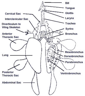
Birds have a respiratory channel terdir top nostril, trakea, bronkus and tuberculosis-apru. At the bottom of the trakea there is a sound tool called siring. Birds have a tool called bantu respiratory sac air with lungs. Function of the air sac, among others, to assist respiratory and help raise siring cavity so that it can strengthen the voice. Respiratory process occurs in birds as follows. If berkontaksi rib muscles, rib to move forward and to the sternum to move down. Thoracic cavity and a large tekanannya decreased. This causes the air into the lungs and then into the air sac. In the rib muscles become weak, broken bones to move back to the breastbone and moving toward the top. Thoracic cavity and wane tekanannya become large, causing the air out of the lungs. Similarly from the air sac air out through the paru-apru. Of oxygen by the lungs occurred at the time of inspiration and ekspirasi. Exchange of gas occurs only in the lungs.

Reptiles
Reptiles breathe with lungs. The oxygen and carbon dioxide expenditure occurred in the lungs. Exit and entry of air from the lungs out because of the movements of the rib. Channel consists of respiratory smellers, trakea, bronkus and lungs.

Frog
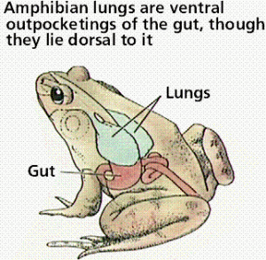
Frog life cycle in the form of changes or metamorfosis. At the time a young tadpole and adult life in the land. At nula with gill breathing tadpole that is outside on the back of the head. Gill always vibrates in the water around the gill always change. Terlarut the oxygen in the water berdifusi in the capillary blood vessel in the gill.
After some time outside the gill is a gill akan change in the way terbentuknya skin fold from the front to the back of the gill cover so that the outside. Adult frog lives on land, with pernapasannya lungs. In addition to the lungs, oxygen can berdifusi in the mouth cavity through the peritoneal cavity through the mouth and skin.

Fish
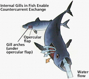
Gold fish with gill breathing in the left and right side head. Each have four closed gill by gill cover (operkulum). Respiratory process in fish is a way to open and close the mouth alternately with opening and closing the gill cover. At the time the mouth opens, the water into the mouth cavity while closing the gill cover. Terlarut the oxygen in the water to enter berdifusi in capillary blood vessel in the gill. And at the time of close, close the gill opening and the water out of the mouth cavity through the gill. Together with the discharge of water through the gill, carbon dioxide is removed. Exchange of oxygen and carbon dioxide occurs in the gill sheet.
Insects have sitem respiratory system called trakea. Oxygen needed by the body's cells to oxidation is not distributed by blood but distributed by the trakea ramify throughout the body. Small branches that penetrate the network trakea body called trakeolus. Respiratory air to the entrance through the mouth but not through the stigma (spirakel).
Respiratory process in the insect occur as follows. With the contraction of the body musculature, the insect's body and into the mengempis regularly. In the body of the insect, the air entering through the stigma, to the next entry in trakea, and then into trakeolus and finally into the body cells. Berdifusi oxygen to cells in the body. Carbon dioxide is removed through the respiratory system trakea also issued through the end stigma at the time of insect bodies mengempis.

Soil worm
Earthworms do not have any special respiratory equipment. Skin contains a lot of mucus gland, the body so that skin becomes wet and humid. Oxygen is needed by the body go through the entire body surface diffusion. Expenditure of carbon dioxide through the body surface.

Protozoa
Protozoa not have special respiratory equipment to obtain oxygen and remove carbon dioxide. Oxygen into the cell plasma membrane malalui the diffusion. Similarly carbon dioxide from the cells in deikeluarkan through the plasma membrane.


====================================================================


Versi Indonesia


Sistem Pernapasan Hewan


Burung
Burung mempunyai saluran pernapasan yang terdir atas lubang hidung, trakea, bronkus dan paru-apru. Pada bagian bawah trakea terdapat alat suara disebut siring. Burung mempunyai alat bantu pernapasan yang disebut pundi-pundi udara yang berhubungan dengan paru-paru. Fungsi pundi-pundi udara antara lain untuk membantu pernapasan dan membantu membesarkan rongga siring sehingga dapat memperkeras suara. Proses pernapasan pada burung terjadi sebagai berikut. Jika otot tulang rusuk berkontaksi, tulang rusuk bergerak ke arah depan dan tulang dada bergerak ke bawah. Rongga dada menjadi besar dan tekanannya menurun. Hal ini menyebabkan udara masuk ke dalam paru-paru dan selanjutnya masuk ke dalam pundi-pundi udara. Pada waktu otot tulang rusuk mengendur, tulang rusak bergerak ke arah belakang dan tulang dada bergerak ke arah atas. Rongga dada mengecil dan tekanannya menjadi besar, mengakibatkan udara keluar dari paru-paru. Demikian juga udara dari pundi-pundi udara keluar melalui paru-apru. Pengambilan oksigen oleh paru-paru terjadi pada waktu inspirasi dan ekspirasi. Pertukaran gas hanya terjadi di dalam paru-paru.


Pernafasan-burung


Reptil
Reptil bernapas dengan paru-paru. Pengambilan oksigen dan pengeluaran karbondioksida terjadi di dalam paru-paru. Keluar masuknya udara dari dan keluar paru-paru karena adanya gerakan-gerakan dari tulang rusuk. Saluran pernapasan terdiri dari lubang hidung, trakea, bronkus dan paru-paru.

Katak
Katak dalam daur hidupnya mengalami metamorfosis atau perubahan bentuk. Pada waktu muda berupa berudu dan setelah dewasa hidup di darat. Mula-nula berudu bernapas dengan insang luar yang terdapat di bagian belakang kepala. Insang tersebut selalu bergetar yang mengakibatkan air di sekitar insang selalu berganti. Oksigen yang terlarut dalam air berdifusi di dalam pembuluh kapiler darah yang terdapat dalam insang.
Setelah beberapa waktu insang luar ini akan berubah menjadi insang dalam dengan cara terbentuknya lipatan kulit dari arah depan ke belakang sehingga menutupi insang luar. Katak dewasa hidup di darat, pernapasannya dengan paru-paru. Selain dengan paru-paru, oksigen dapat berdifusi dalam rongga mulut yaitu melalui selaput rongga mulut dan juga melalui kulit.


Pernafasan-katak


Ikan
Ikan mas bernapas dengan insang yang terdapat pada sisi kiri dan kanan kepala. Masing-masing mempunyai empat buah insang yang ditutup oleh tutup insang (operkulum). Proses pernapasan pada ikan adalah dengan cara membuka dan menutup mulut secara bergantian dengan membuka dan menutup tutup insang. Pada waktu mulut membuka, air masuk ke dalam rongga mulut sedangkan tutup insang menutup. Oksigen yang terlarut dalam air masuk berdifusi ke dalam pembuluh kapiler darah yang terdapat dalam insang. Dan pada waktu menutup, tutup insang membuka dan air dari rongga mulut keluar melalui insang. Bersamaan dengan keluarnya air melalui insang, karbondioksida dikeluarkan. Pertukaran oksigen dan karbondioksida terjadi pada lembaran insang.
Serangga mempunyai sitem pernapasan yang disebut sistem trakea. Oksigen yang dibutuhkan oleh sel-sel tubuh untuk oksidasi tidak diedarkan oleh darah tetapi diedarkan oleh trakea yang bercabang-cabang ke seluruh tubuh. Cabang kecil trakea yang menembus jaringan tubuh disebut trakeolus. Masuknya udara untuk pernapasan tidak melalui mulut melainkan melalui stigma (spirakel).

Pernafasan-Ikan

Proses pernapasan pada serangga terjadi sebagai berikut. Dengan adanya kontraksi otot-otot tubuh, maka tubuh serangga menjadi mengembang dan mengempis secara teratur. Pada waktu tubuh serangga mengembang, udara masuk melalui stigma, selanjutnya masuk ke dalam trakea, kemudian ke dalam trakeolus dan akhirnya masuk ke dalam sel-sel tubuh. Oksigen berdifusi ke dalam sel-sel tubuh. Karbondioksida hasil pernapasan dikeluarkan melalui sistem trakea juga yang akhirnya dikeluarkan melalui stigma pada waktu tubuh serangga mengempis.

Cacing Tanah
Cacing tanah tidak mempunyai alat pernapasan khusus. Kulitnya banyak mengandung kelenjar lendir, sehingga kulit tubuhnya menjadi basah dan lembab. Oksigen yang diperlukan oleh tubuhnya masuk melalui seluruh permukaan tubuh secara difusi. Pengeluaran karbon dioksida juga melalu permukaan tubuh.

Protozoa
Protozoa tidak mempunyai alat pernapasan khusus untuk memperoleh oksigen dan mengeluarkan karbon dioksida. Oksigen masuk ke dalam sel malalui selaput plasma secara difusi. Demikian juga karbon dioksida dari dalam sel deikeluarkan melalui selaput plasma.

Kelainan/Penyakit pada Pernapasan

DEVIATION / RESPIRATORY DISEASE IN


Respiratory system can experience a variety of interference, either because the system aberration due to respiratory infection or bacteria. Several types of interference are:


-Asma/sesak Breath, the breath due to constriction channel forming smooth muscle wall of the channel berkontraksi hold, due to allergies or lack of hormone adrenalin.


-Asfiksi, interference and the use of oxygen carriage by the network due to sinking, pneumonia, CO poisoned.


-Asidosis, due to increased carbonic acid content and acid in the blood bicarbonate
Adenoid

-face (face stupid), because the breath constringency channel gland swelling limfa (polypus), swelling in the throat (tonsil).


-Pneumonia, pneumonia due to bacterial infection Diplococcus pneumonia.


-Diphtheria, stoppage faring / laring mucus due to infection by the bacteria Corynebacterium diphteriae


-Emfisema, menggelembungnya lung alveolus due to excessive expansion.


-Tuberculosis (TB), lung disease due to infection of the bacteria Mycobacterium tuberculosa.


-Inflammation in the respiratory system:


• bronchitis, inflammation bronkhus.
• laringitis, inflammation laring
• faringitis, inflammation faring
• pleuritis, peritonitis lungs
• renitis, sore nose cavity
• sinusitis, sore at the top of the nose cavity (sinus)


=====================================================


Versi Indonesia

Kelainan/Penyakit pada Pernapasan


Sistem pernapasan dapat mengalami berbagai gangguan, baik karena kelainan sistem pernapasan atau akibat infeksi kuman. Beberapa jenis gangguan antara lain :

-Asma/sesak napas, penyempitan saluran napas akibat otot polos pembentuk dinding saluran terus berkontraksi, disebabkan alergi atau kekurangan hormon adrenalin.

-Asfiksi, gangguan pengangkutan dan penggunaan oksigen oleh jaringan akibat tenggelam, pneumonia, keracunan CO.

-Asidosis, akibat peningkatan kadar asam karbonat dan asam bikarbonat dalam darah
-Wajah adenoid (wajah bodoh), penyempitan saluran napas karena pembengkakan kelenjar limfa (polip), pembengkakan di tekak (amandel).

-Pneumonia, radang paru-paru akibat infeksi bakteri Diplococcus pneumonia.

-Difteri, penyumbatan faring/laring oleh lendir akibat infeksi bakteri Corynebacterium diphteriae
-Emfisema, menggelembungnya paru-paru akibat perluasan alveolus berlebihan.

-Tuberculosis (TBC), penyakit paru-paru akibat infeksi bakteri Mycobacterium tuberculosa.

-Peradangan pada sistem pernapasan :

  • bronchitis, radang bronkhus.
  • laringitis, radang laring
  • faringitis, radang faring
  • pleuritis, radang selaput paru-paru
  • renitis, radang rongga hidung
  • sinusitis, radang pada bagian atas rongga hidung (sinus)

macam-macam pernafasan

TYPES OF RESPIRATORY


1. Respiratory chest
Respiratory chest held in 2 stages, namely:

Inspiration, occurs when the muscles between the rib outside berkontraksi, rib up, volume up chest cavity, lungs expand, so the air pressure becomes smaller from the air atmosphere, so that the incoming air.

Ekspirasi, occurs when the muscles between the rib outside berelaksasi, rib akan initially attracted to the position, the volume of off thoracic cavity, chest cavity air pressure increases, air pressure in the lungs is higher than the air atmosphere, a result the air out.


2. Respiratory stomach
Respiratory stomach held in two phases, namely:
Inspiration, occurs when the diaphragm muscle berkontraksi, horizontal diaphragm cause chest cavity volume increase air pressure so that the decline followed the lungs causing the air pressure lower than the air pressure and air entering the atmosphere.

Ekspirasi, beginning with the muscles and diaphragm berelaksasi stomach muscle wall berkontraksi the diaphragm up and vaulted the thoracic cavity, chest cavity so that the volume decline and tekanannya increased so that the air in the lungs out. Respiratory generally occurs when the stomach to sleep.

GAS Exchange Mechanism
Transportation O2

Gas exchange between O2 with CO2 occurs in the alveolus and the body of the network, through a process of diffusion. Until the oxygen in the alveolus akan berdifusi penetrate membrane alveolus and berikatan with haemoglobin (Hb) in the blood called deoksigenasi and compound oksihemoglobin (HBO) as a reaction to the following:

Approximately 97% oxygen in the form of a compound oksihemoglobin, only 2 - 3%, which is dissolved in blood plasma will be carried by blood to the entire body of the network, and the next release will be a diffusion of oxygen from blood to the body of the network,

Transport CO2

Carbon dioxide (CO2) produced from the process of cell respiration akan berdifusi in the blood which will then be transported to the lungs for respiratory air as issued.

There are three (3) how transportation CO2:

Sebagai ion karbonat (HCO3), sekitar 60 – 70%.

Sebagai karbominohemoglobin (HbCO2), sekitar 25%

Sebagai asam karbonat (H2CO3) sekitar 6 – 10%.


==============================================================


Versi Imdonesi


MACAM – MACAM PERNAPASAN


1. Pernapasan Dada

Pernapasan dada berlangsung dalam 2 tahap, yaitu :

Inspirasi, terjadi bila otot antar tulang rusuk luar berkontraksi, tulang rusuk terangkat, volume rongga dada membesar, paru-paru mengembang, sehingga tekanan udaranya menjadi lebih kecil dari udara atmosfer, sehingga udara masuk.

Ekspirasi, terjadi bila otot antar tulang rusuk luar berelaksasi, tulang rusuk akan tertarik ke posisi semula, volume rongga dada mengecil, tekanan udara rongga dada meningkat, tekanan udara dalam paru-paru lebih tinggi dari udara atmosfer, akibatnya udara keluar.


2. Pernapasan Perut

Pernapasan perut berlangsung dalam dua tahap, yaitu :

Inspirasi, terjadi bila otot diafragma berkontraksi, diafragma mendatar mengakibatkan volume rongga dada membesar sehingga tekanan udaranya mengecil dan diikuti paru-paru yang mengembang mengakibatkan tekanan udaranya lebih kecil dari tekanan udara atmosfer dan udara masuk.

Ekspirasi, diawali dengan otot diafragma berelaksasi dan otot dinding perut berkontraksi menyebabkan diafragma terangkat dan melengkung menekan rongga dada, sehingga volume rongga dada mengecil dan tekanannya meningkat sehingga udara dalam paru-paru keluar. Pernapasan perut umumnya terjadi saat tidur.

MEKANISME PERTUKARAN GAS

Pengangkutan O2

Pertukaran gas antara O2 dengan CO2 terjadi di dalam alveolus dan jaringan tubuh, melalui proses difusi. Oksigen yang sampai di alveolus akan berdifusi menembus selaput alveolus dan berikatan dengan haemoglobin (Hb) dalam darah yang disebut deoksigenasi dan menghasilkan senyawa oksihemoglobin (HbO) seperti reaksi berikut :

Sekitar 97% oksigen dalam bentuk senyawa oksihemoglobin, hanya 2 – 3% yang larut dalam plasma darah akan dibawa oleh darah ke seluruh jaringan tubuh, dan selanjutnya akan terjadi pelepasan oksigen secara difusi dari darah ke jaringan tubuh,

Pengangkutan CO2

Karbondioksida (CO2) yang dihasilkan dari proses respirasi sel akan berdifusi ke dalam darah yang selanjutnya akan diangkut ke paru-paru untu

k dikeluarkan sebagai udara pernapasan.

Ada 3 (tiga) cara pengangkutan CO2 :

Sebagai ion karbonat (HCO3), sekitar 60 – 70%.

Sebagai karbominohemoglobin (HbCO2), sekitar 25%

Sebagai asam karbonat (H2CO3) sekitar 6 – 10%.

Friday, May 22, 2009

anatomi tubuh manusia dan hewan

A N A T O M I


anatomi tubuh manusia

Terlebih dahulu saya ingin mengenalkan diri saya.
Langsung saja ya… nama saya zulfan hamdi. Saya tamatan smu hangtuah belawan, pekerjaan saya pengangguran, dll
Udah ya mpek situ saja perkenalan saya..

Disini saya mencoba buat blog dalam dua versi bahasa. Yaitu Indonesia dan English.
Sekarang kita mulai pembahasan dari versi english duluan ya

Baiklah teman-teman semua dan bagi para pembaca blog ini.
Saya akan mulai menerangkan apa itu anatomi tubuh manusia dan hewan. Kalau ada kesalahan mohon di maaf kan. (saya juga masih belajar)
Masuk ke inti masalah yaa..

English Version
Anatomy (derived from the Greek anatomia ἀνατομία, from ἀνατέμνειν anatemnein, which means that the cut) is a branch of biology dealing with the structure and organization of living things. There are also zootomi or animal anatomy and plant anatomy or fitotomi. Some of the science of anatomy is a branch of comparative anatomy, histology, and human anatomy.

Anatomy Animals
Anatomy of animals also known as comparative anatomy or morphology if the animals learn the structure of various animals, and the anatomy is called special if only learn one type of animal only.

Human Anatomy
View from the point of usefulness, the most important part of the anatomy that is special about human learning with the various different approaches. From the point of medical, anatomy consists of a variety of forms of knowledge, disposition, size, structure and relationships of various human body healthy, so often referred to as descriptive or topographical anatomy. The complexity of the human body there are only a few human anatomy expert professionals who really take this field of science; most have specialized in a specific part such as the brain or in part.
Topographic anatomy should be learned with the surgery and repeated examination in the human body that has died ([[kadaver]) Anatomy of a science is not common, but should really have a high accuracy because it can be used in emergency situations. Pathology is the science of the anatomy of organs and that have a difference in the illness. This knowledge is applied for various purposes such as surgery and gynecology.


Versi Indonesia
Anatomi (berasal dari bahasa Yunani ἀνατομία anatomia, dari ἀνατέμνειν anatemnein, yang berarti memotong) adalah cabang dari biologi yang berhubungan dengan struktur dan organisasi dari makhluk hidup. Terdapat juga anatomi hewan atau zootomi dan anatomi tumbuhan atau fitotomi. Beberapa cabang ilmu anatomi adalah anatomi perbandingan, histologi, dan anatomi manusia.

Anatomi Hewan
Anatomi hewan juga disebut sebagai anatomi perbandingan atau morfologi hewan jika mempelajari struktur berbagai hewan, dan disebut anatomi khusus jika hanya mempelajari satu jenis hewan saja.

Anatomi Manusia
Dilihat dari sudut kegunaan, bagian paling penting dari anatomi khusus adalah yang mempelajari tentang manusia dengan berbagai macam pendekatan yang berbeda. Dari sudut medis, anatomi terdiri dari berbagai pengetahuan tentang bentuk, letak, ukuran, dan hubungan berbagai struktur dari tubuh manusia sehat sehingga sering disebut sebagai anatomi deskriptif atau topografis. Kerumitan tubuh manusia menyebabkan hanya ada sedikit ahli anatomi manusia profesional yang benar-benar menguasai bidang ilmu ini; sebagian besar memiliki spesialisasi di bagian tertentu seperti otak atau bagian dalam.

Anatomi topografi harus dipelajari dengan pembedahan dan pemeriksaan berulang kali pada tubuh manusia yang telah meninggal ([[kadaver]) Anatomi bukan sekedar ilmu biasa, namun harus benar-benar mempunyai keakuratan yang tinggi karena dapat digunakan dalam situasi yang darurat. Patologi anatomi adalah ilmu mengenai organ yang memiliki kelainan dan dalam keadaan sakit. Ilmu ini diterapkan untuk berbagai tujuan seperti bedah dan ginekologi.